Introduction:
The term “pes planus” is a broad category of pedal complaints that is a descriptive term rather than an actual diagnosis. This term simply means flat foot, which does not provide the podiatrists and foot and ankle surgeons an appreciation of the broad range of complaints and difficulties that may be associated with this pathological entity. Pes planus presents in
all age groups and is often described by a range of names including pes planus, pes planovalgus, pes valgus, flatfoot, peritalar subluxation, and several others.
Biomechanics:
The foot is a complex organ, which is composed of many interacting anatomical components in a dynamic manner. As a result of these interactions the foot functions in a multiplanar manner. Flatfoot exists in any situation in which the medial longitudinal arch of the foot is flat or collapsed and in pathological situations.
Posterior Tibial Tendon Dysfunction
Etiology:
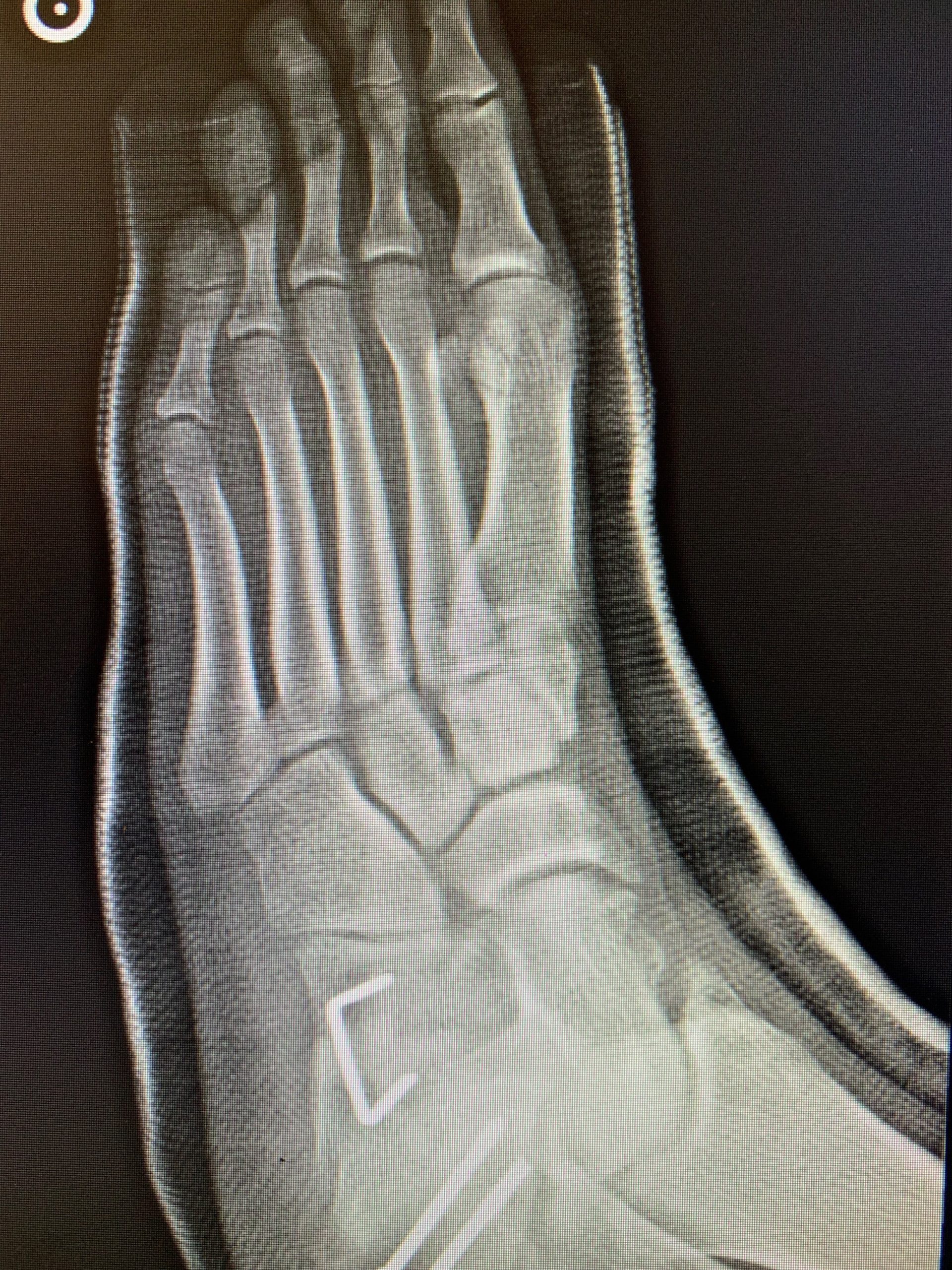
Posterior tibial tendon dysfunction is referred to by various terms and one of these, adult acquired flatfoot, is quite appropriate as this disorder occurs in adults rather than children. The posterior tibialis tendon can rupture, becomes attenuated, or fails to function the mechanisms of supporting the medial arch and keeping the heel in neutral position.
Epidemiology: in all studies it show a strong predilection for females over males, 4:1 female to Adult Acquired Flat Feet occurs most commonly in patients over 50 years of age . A
study by Holmes and Mann found 76% of patients were women and 52% had diabetes, obesity, or hypertension. It is difficult to determine if these factors are correlated or actual causes as all three characteristics are more common in patients of advancing age. A preexisting flatfoot deformity is thought to exist in almost 100% of patients. These statistics are very similar to the patients that we see at our podiatry offices in Chino and Upland. Our foot doctors in Chino and Upland can correlate very well with these figures.
Examination:
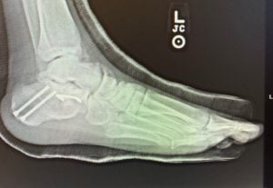
Our podiatrists at Empire Foot and Ankle Center in both Upland and Chino locations are well trained and experienced to treat adult flat feet. Our foot and ankle specialists will perform a complete physical examination after obtaining medical history from the patients to come up with the best assessment and treatment plan for the patients in Inland Empire with flat foot deformity. Our foot and ankle surgeons use the newest diagnostic modalities including one of the most modern portable and wireless XRAY machines and an ultrasound machine to come up with proper diagnosis and treatment plan in both our Chino and Upland locations. Our upland foot doctors and Chino podiatrists come up with a unique treatment plan for each patient to relieve any foot and ankle pain and discomfort as soon as possible.
Treatment:
There are both non-surgical and surgical treatments for this pathology and podiatrists at Empire Foot and Ankle are well trained to treat patients either way. Early stages of posterior tibial tendon dysfunction may be treated relatively conservatively, including rest, ice, anti-inflammatories, and weightbearing immobilization with a below knee cast or cast
boot/cam walker for 6-8 weeks. At this point the patient is re-evaluated at our state of art Chino and Upland offices and further treatment is determined. If no deformity progression is seen the patient may be started on a physical therapy regimen and placed in a custom foot orthotic or ankle foot orthotic. If the deformity has progressed or presented as a progressive stage considerations for foot and ankle surgery may be necessary. Our foot and ankle surgeons will not perform any surgery unless they have exhausted all conservative treatments.
Please note that there is not only one kind of surgery for this pathology and there are different surgical procedures depending on the severity and plane dominance for this pathology.
Recommendation:
If you are suffering from flat foot deformity, please do not wait and come see one of our podiatrists in Chino or Upland location. Flat foot can progress and worsen over time but with early conservative therapy, many patients return back to activity and do not require any surgery in the future.
Our podiatry offices are within few miles from the cities of Upland, Chino, Ontario, Chino Hills, Montclair, Rancho Cucamonga, Fontana, Pomona, Covina, Claremont, San Dimas, Eastvale, Alta Loma. Our expert podiatrists and foot and ankle surgeons are well trained to perform conservative and surgical treatments for any foot and ankle diseases including flat feet, bunions, limb salvage, diabetic foot care, ingrown nail, fungus, plantar fasciitis, pediatric foot deformities, hammertoes, diabetic foot wounds, rheumatoid arthritis, wart, corns and calluses, circulatory system diseases, foot fractures, ankle fractures.
References:
Akcali O, et al. Effects of Lower Extremity Rotation on Prognosis of Flexible Flatfoot in Children. Foot & AnkleInternational, Sept 2000; 21(9): 772-774.
Baylis WJ and Rzonca EC. Functional and Structural Limb Length Discrepancies: Evaluation and Treatment.Clinics in Podiatric Medicine and Surgery, July 1988; 5(3): 509-520.
Benard MA. Treatment of Skewfoot by Multiple Lesser Tarsal Osteotomies and Calcaneal Osteotomy. The
Journal of Foot Surgery, 1990; 29(5): 504-509.
Bouysset M, et al. Posterior Tibial Tendon and Subtalar Joint Complex in Rheumatoid Arthritis: Magnetic
Resonance Imaging Study. The Journal of Rheumatology, 2003; 30(9): 1951-1954.
Brown, PB. Rheumatoid Flatfoot. Journal of the American Podiatric Medical Association, Jan 1987; 77(1): 39-41.
Cappello T and Mosca VS. Metatarsus Adductus and Skewfoot. Foot and Ankle Clinics, Dec 1998; 3(4): 683-700.
Cass AD and Camasta CA. A Review of Tarsal Coalition and Pes Planovalgus: Clinical Examination, Diagnostic
Imaging, and Surgical Planning. The Journal of Foot and Ankle Surgery, 2010; 49: 274-293.
Chu IT, et al. Experimental Flatfoot Model: The Contribution of Dynamic Loading. Foot and Ankle International,
Mar 2001; 22(3): 220-225.
Coughlin MJ. The Rheumatoid Foot. Postgraduate Medicine, Apr 1984; 75(5): 207-216.
Deland, et al. Adult Acquired Flatfoot Deformity at the Talonavicular Joint: Reconstruction of the Spring
Ligament in an In Vitro Model. Foot and Ankle, July 1992; 13(6): 327-332.
Dellon AL and Barrett SL. Sinus Tarsi Denervation: Clinical Results, Mar 2005; 95(2): 108-113.
Downey MS. Tarsal Coalition. In McGlamary’s Comprehensive Textbook of Foot and Ankle Surgery. Vol 1, 3rd ed.
Lippincott Williams & Wilkins; 2001: 993-1031.
Dyal CM, et al. Pes Planus in pateints with Posterior Tibial Tendon Insufficiency: Asymptomatic Versus
Symptomatic Foot. Foot and Ankle International, Feb 1997; 18(2): 85-88.
Evans D. Calcaneo-Valgus Deformity. The Journal of Bone and Joint Surgery, Aug 1975; 57-B(3):270-278.
Geist ES. The Accessory Scaphoid Bone. The Journal of Bone and Joint Surgery, 1925; 7: 570-574.
GogiaPP and Braatz JH. Validity and reliability of leg length measurements. Journal of Orthopedic and Sports
Physical Therapy, Oct 1986; 8(4): 185-188.Feb 1984; 74(2): 98-103.
Gokce M, et al. Down syndrome: orthopedic issues. Current Opinion in Pediatrics, Feb 2008; 20(1): 30-36.
Green DL and Carol A. Planal Dominance. Journal of the American Podiatry Association, Feb 1984; 74(2): 98-103.
Grogan DP, et al. The Painful Accessory Navicular: A Clinical and Histopathological Study. Foot and Ankle, Dec
1989; 10(3): 164-169.
Grumbine NA, et al. Calcaneal “L” Osteotomy: A Retrospective Study, 1991; 30(4): 325-335.
Gurney B. Leg length discrepancy. Gait and Posture, 2002; 15: 195-206.
Hagmann S, et al. Skewfoot. Foot and Ankle Clinics of North America, Sep 2009; 14(3): 409-434.
Harris E. Pediatric Neurology in Introduction to Podopediatrics, 2nd edition. Ed. Thompson P and Volpe R 2001:121-160.
Harris RI and Beath T. Etiology of Peroneal Spastic Flat Foot. The Journal of Bone and Joint Surgery, Nov 1948;30B(4): 624-634.Helal B. Cobb Repair for Tibialis Posterior Tendon Rupture. Journal of Foot and Ankle Surgery, 1990; 29(4): 349-
352.
Heyman CH, et al. Mobilization of the Tarsometatarsal and Intermetatarsal Joints for the Correction of Resistant
Adduction of the Fore Part of the Foot in Congenital Club-Foot or Congenital Metatarsus Varus. The Journal of bone and Joint Surgery, Apr 1958; 40-A(2): 299-310.
Hoke M. An Operation for the Correction of Extremely Relaxed Flat Feet. The Journal of Bone and Joint Surgery,Ap 1931; 13: 773-783.
Holmes GB and Mann RA. Possible Epidemiological Factors Asscoatied with Rupture of the Posterior Tibial
Tendon. Foot and Ankle International, Feb 1992; 13(2): 70-79.
Hubbard AM, et al. Magnetic Resonance Imaging of Skewfoot. The Journal of Bone and Joint Surgery, Mar 1996;78-A (3): 389-397.
Hutchinson B. Pediatric Metatarsus Adductus and Skewfoot Deformity. Clinics in Podiatric Medicine and Surgery, Jan 2010; 27(1): 93-104.
Jennings MM and Christensen JC. The Effects of Sectioning the Spring Ligament on Rearfoot Stability and
Posterior Tibial Tendon Efficiency. The Journal of Foot and Ankle Surgery, May 2oo8; 47(3): 219-224.
Johnson JB. A Preliminary Report on Chondrotomies: A New Surgical Approach for Metatarsus Adductus inChildren. Journal of the American Podiatry Association, Dec 1978; 68(12): 808-813.
Johnson KA and Strom DE. Tibialis Posterior Tendon Dysfunction. Clinical Orthopedics and Related Research, Feb
1989; 239: 196-206.
Kawashima T and Uhthoff HK. Prenatal Development Around the Sustentaculum Tali and Its Relation toTalocalcaneal Coalitions. Journal of Pediatric Orthopedics, 1990; 10(2): 238-243.
Kernabach KJ, et al. Bilateral Single-Stage Middle Facet Talocalcaneal Resection Combined with Flatfoot
Reconstruction: A Report of 3 Cases and Review of the Literature. Investigations Involving Middle Facet Coalitions – Part 1. Journal of Foot and Ankle Surgery, May 2008; 47(3): 180-190.
Kidner FC. The Prehallux (Accessory Scaphoid) In Its Relation To Flat-Foot. The Journal of Bone and Joint Surgery, 1929; 11: 831-837.
Knupp M, et al. Triple Arthrodesis in Rheumatoid Arthritis. Foot and Ankle International, Mar 2008; 29(3): 293-297.
Koutsogiannis E. Treatment of Mobile Flat Foot By Displacement Osteotomy of the Calcaneus. The Journal of Bone and Joint Surgery, Feb 1971; 53B(1): 96-100. Kumai T, et al. Histopathological Studyof Nonosseous Tarsal Coalition. Foot and Ankle International, Aug 1998;19(8): 525-531.
Kuwada GT. Long-Term Retrospective Analysis of the Treatment of Sinus Tarsi Syndrome. The Journal of Foot and Ankle Surgery, Jan 1994; 33(1): 28-29.
Labovitz JM. The Algorithmic Approach to Pediatric Flexible Pes Planovalgus. Clinics in Podiatric Medicine and Surgery, Jan 2006; 23(1): 57-76.
Lapidus PW. A Quarter of a Century of Experience With the Operative Correction of the Metatarsus Varus Primus in Hallux Valgus. Bulletin of Hospital Joint Diseases, Oct 1956; 17(2): 404-421.
Lord JP. Correction of Extreme Flatfoot. Value of Osteotomy of Os Calcis and Inward Displacement of Posterior
Fragment (Gleich Operation). Journal of the American Medical Association, Nov 1923; 81: 1502-1507.
Lowman CL. An Operative Method for correction of Certain Forms of Flatfoot. Journal of the American Medical Association, Nov 1923; 81: 1500-1502.
Lowy LL. Pediatric Peroneal Spastic Flatfoot in the Absence of Coalition: A Suggested Protocol. Journal of the American Podiatric Medical Association, Ap 1998; 88(4): 181-191.
Maenpaa, H, et al. Why do Ankle Arthrodeses Fail in Patients with Rheumatic Disease? Foot and Ankle International, May 2001; 22(5): 403-408.
Mann RA and Thompson FM. Rupture of the posterior tibial tendon causing flat foot. Surgical treatment. Journal of Bone and Joint Surgery, Apr 1985;67-A(4): 556-561.
Masterson E, et al. The Planovalgus Rheumatoid Foot – Is Tibialis Posterior Tendon Rupture A Factor? British Journal of Rheumatology, 1995; 34(7): 645-646.
Minaker K and Little H. Painful feet in rheumatoid arthritis. Canadian Medical Journal, Oct 1973; 109: 724-730. Mosca VS. Calcaneal Lengthening for Valgus Deformity of the Hindfoot. Results in Children Who Had Severe, Symptomatic Flatfoot and Skewfoot. Journal of Bone and Joint Surgery, Apr 1995; 77-A(4): 500-512.
Mueller T. Acquired Flatfoot Secondary to Tibialis Posterior Dysfunction: Biomechanical Aspects. The Journal of Foot Surgery, 1991; 30(1): 2-11.
Myerson MS. Adult Acquired Flatfoot Deformity: Treatment of Dysfunction of the Posterior Tibial Tendon. The
Journal of Bone and Joint Surgery, May 1996; 78-A(5):780-792.
Myerson MS, et al. Treatment of Stage II Posterior Tibial Tendon Deficiency With Flexor Digitorum Longus Tendon Transfer and Calcaneal Osteotomy. Foot and Ankle International, July 2004; 25(7): 445-450.
Napiontek M. Skewfoot. Journal of Pediatric Orthopedics, 2002; 22(1): 130-133.
Niki H, et al. The Effect of Posterior Tibial Tendon Dysfunction on Hindfoot Kinematics. Foot and Ankle International, Apr 2001; 22(40): 292-300.
Nittaya L, et al. Tarsal Sinus: Arthrographic, MR Imaging, MR Arthrographic, and Pathologic Findings in Cadavers and Retrospective Study Data in Patients with Sinus Tarsi Syndrome. Radiology, 2001; 219(3): 802-810.
Oloff LM, et al. Subtalar Joint Arthroscopy for Sinus Tarsi Syndrome: A Review of 29 Cases. The Journal of Foot and Ankle Surgery, May 2001; 40(3): 152-157.
Osborn PM. Quiz case. European Journal of Radiology,2003; 47: 60-63.
Pasani G, et al. Sinus tarsi syndrome and subtalar joint instability. Clinics in Podiatric Medicine and Surgery, Jan 2005; 22(1): 63-77. Perlman MD and Wertheimer SJ. Tarsal Coalitions. The Journal of Foot Surgery, 1986: 25(1): 58-67.
Prasher VP, et al. Podiatric disorders Among Children with Down Syndrome and Learning Disability. Developmental Medicine and Child Neurology, 1995; 37: 131-134.
Prichasuk S, et al. Kidner Procedure for Symptomatic Accessory Navicular and Its Relation to Pes Planus. Foot and Ankle International, Aug 1995; 16(8): 500-503.
Rankin EA and Baker GI. Rigid Flatfoot in the Young Adult. Clinical Orthopedics and Related Research, Oct 1974;104: 244-248.
Root ML, et al. Forces Acting on the Foot During Propulsion (Ch. 7) in normal and abnormal Function of the Foot Vol 2, Clinical Biomechanics Corporation, 1977.
Sabharwal S. Current Concepts Review: Blount Disease. Journal of Bone and Joint Surgery, July 2009; 91-A(7): 1758-1776.
Saxena A and Erickson S. Tarsal Coalitions: Activity Levels With and Without Surgery. Journal of the American Podiatric Medical Association, Aug 2003; 93(4): 259-263.
Schweinberger MH and Roukis TS. Soft Tissue Correction of Ankle Equinus Contracture. Clinics in Podiatric Medicine and Surgery, Oct 2008; 25(4): 571-585.
Sella EJ., et al. Biomechanics of the Accessory Navicular Synchondrosis. Foot and Ankle, Dec 1987; 8(3): 156-163.
Scott AT and Tuten HR. Calcaneonavicular Coalition Resection with Extensor Digitorum Brevis Interposition in Adults. Foot and Ankle International, Aug 2007; 28(8): 890-895.
Shirley ED and Sponseller PD. Marfan Syndrome. Journal of the American Academy of Orthopedic Surgeons, Sept 2009; 17(9): 572-581.
Silver, CM, et al. Calcaneal Osteotomy for Valgus and Varus Deformities of the Foot in Cerebral Palsy: A Preliminary Report of Twenty-Seven Operations. The Journal of Bone and Joint Surgery, Mar 1967; 49A (2): 232-
246.
Safiropoulos G, et al. Flat foot and femoral anteversion in children – A prospective study. The Foot, 2009; 19:50-54.
Ugolini PA and Raikin SM. The accessory navicular. Foot and Ankle Clinics of North America, 2004; 9: 165-180.
Wan SC. Metatarsus Adductus and Skewfoot Deformity. Clinics in Podiatric Medicine and Surgery, Jan 2006; 23(1): 21-40.
Whisler RL, et al. Rheumatology, A Clinical Overview. Clinics in Podiatric Medicine and Surgery, Jan 2002; 19(1):149-161.
Woodburn J, et al. Looking through the ‘window of opportunity’: is there a new paradigm of podiatry care on the horizon in early rheumatoid arthritis? Journal of Foot and Ankle Research, 2010; 3(8): 1-10.
Woodburn J, et al. Changes in 3D Joint Kinematics Support the Continuous Use of Orthoses in the Management of Painful Rearfoot Deformity in Rheumatoid Arthritis. The Journal of Rheumatology, 2003; 30(11): 2356-2364.
Woodburn J, et al. A Randomized Controlled Trial of Foot Orthoses in Rheumatoid Arthritis. The Journal of Rheumatology, 2002; 29(7): 1377-1383.